


Obstetrics
In the field of obstetrics, we offer our patients preparation for pregnancy and its management from the diagnosis of pregnancy to delivery. We provide careful care based on the highest standards of the Polish Society of Gynecology and Obstetricians, we carefully evaluate the test results and perform a control ultrasound.
Obstetrics
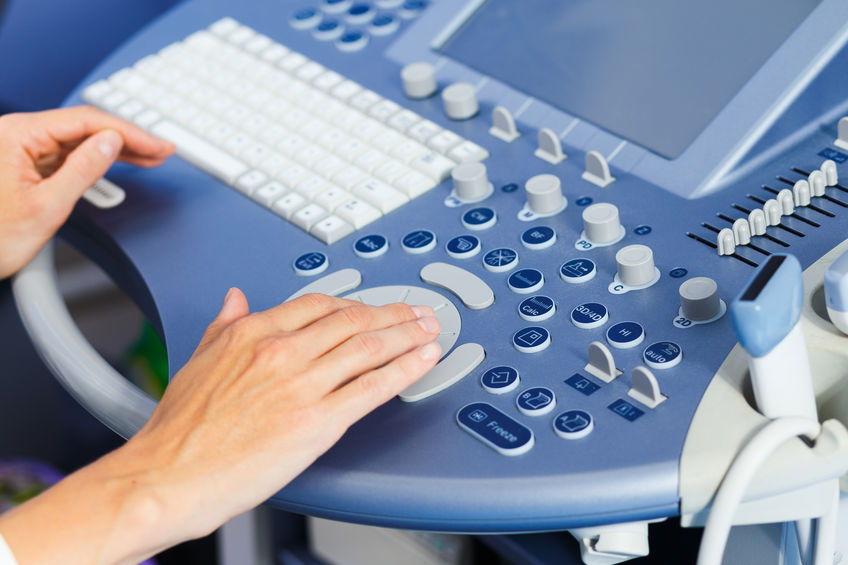
We work on the best quality, regularly serviced equipment for diagnostic imaging. Obstetric ultrasound 2/3 / 4D is performed in our offices. Tests are carried out in warm, soundproofed offices. Each patient can use the intimate hygiene cabin, she is provided with disposable slippers and a skirt. We make every effort to ensure that the visit in our offices is comfortable and the patient feels confident and at ease – which is confirmed by the Safe Office Certificate issued to us.
On a daily basis, we cooperate with outstanding specialists (including in the field of prenatal examinations), with whom we consult sudden and complicated cases, giving the patient a sense of security. Some additional examinations are performed outside our offices in cooperation with the best specialists.
Vaginal (transvaginal) ultrasound
In early pregnancy, it is the basic test to confirm it. It allows to visualize the embryo already in the 4th week of pregnancy, the heart rate of the embryo from the 6th week of pregnancy. During pregnancy, vaginal ultrasound is performed until the 14th week.
With this test, the length of the cervix, the structure of the uterine body and the structure of the ovaries can be assessed.
Ultrasound of the fetus through the abdominal wall
It is performed in pregnant women to assess the fetus at 11-14 weeks of pregnancy (genetic USG), after 20 weeks of pregnancy (prenatal USG) and after 30 weeks of pregnancy. The ultrasound is performed in the supine position. The doctor covers the patient’s skin with gel, then moves the head of the apparatus over the uterus. The image showing the various parts of the fetus’s body and organs is visible on the monitor and recorded on paper.
This examination takes approximately 20 – 30 minutes.
2D ultrasound
The basic part of the pregnancy ultrasound examination is the presentation of a 2D image, where we have the opportunity to observe the cross-section of the inside of the uterus and the general outline of the fetal body with the image of the developing internal organs. Thanks to this, it is possible to assess the size and shape of various parts of the fetal body (mainly internal organs, i.e. the heart, kidneys, brain, stomach, bladder), its structure, as well as the placenta and the amount of amniotic fluid. Importantly, in this study we only get flat sections of the fetal body, which is the main difference between it and the 3D and 4D examination.
Using the ultrasound machine, we can perform a Doppler examination, which allows the assessment of blood flow through the vessels of the fetus and mother thanks to color ultrasound, which allows for obtaining knowledge about the functions of the heart and other organs.
3D ultrasound
These devices enable three-dimensional reconstruction of the image of the fetus (3D) and the inside of the uterus. As a result of the analysis of the 3D view, a precise and clear image of the child is obtained, which is easy for anyone to read. Meanwhile, in a 2D image, very often only a doctor is able to distinguish individual parts of the fetus. This image also gives anatomical sections, unavailable in simpler studies, helpful in more detailed analyzes.
4D ultrasound
It is three-dimensional imaging in real time, allowing the observation of fetal movements and the work of the heart. It allows for more in-depth observation of the child – for example, determining the order in which the specific motor skills of the fetus appear.
4D examination is more readable for parents than two-dimensional cross-sections, but only 4D ultrasound means registering a three-dimensional image in motion. A two-dimensional study is a flat cross-section, a 3D study is a static image in three dimensions, while a 4D study is a moving three-dimensional image. This technology allows for precise evaluation of the brain, spine, heart, kidneys and other organs.

Prenatal testing
Prenatal tests are aimed at detecting genetic diseases, developmental and cardiological defects in the fetus at an early stage of pregnancy. They enable a quick response, establish a treatment plan during pregnancy or after delivery, and help future parents to get used to the possible consequences of a diagnosed disease.
Any woman expecting a baby can undergo prenatal tests. However, there is a group of women who should perform them compulsorily. The obligatory prenatal tests are indicated: age above 35 years of age, a history of genetic or metabolic disease (in a child or in a family). This group of women is covered by a free prenatal testing program reimbursed by the National Health Fund.
School of childbirth
An important element during pregnancy is preparation for childbirth and caring for your baby. We encourage all couples expecting children to participate in birth schools. During the meetings, professional preparation for childbirth is ensured, the stages of labor, methods of pain relief and preparation for the puerperium are discussed. Practical activities include caring for a newborn baby, breastfeeding, bathing a newborn baby and much more. Most Birthing Schools offer advice from a certified lactation advisor, CTG records and patronage visits by a midwife – environmental.